In this article we will discuss about Trypanosoma Gambiense:- 1. Historical Background of Trypanosoma Gambiense 2. Distribution of Trypanosoma Gambiense 3. Habit and Habitat 4. Structure 5. Polymorphic Forms 6. Location 7. Nutrition 8. Respiration 9. Excretion 10. Reproduction 11. Life Cycle 12. Transmission 13. Reservoirs.
Contents:
- Historical Background of Trypanosoma Gambiense
- Distribution of Trypanosoma Gambiense
- Habit and Habitat of Trypanosoma Gambiense
- Structure of Trypanosoma Gambiense
- Polymorphic Forms of Trypanosoma Gambiense
- Location of Trypanosoma Gambiense
- Nutrition in Trypanosoma Gambiense
- Respiration in Trypanosoma Gambiense
- Excretion in Trypanosoma Gambiense
- Reproduction in Trypanosoma Gambiense
- Life Cycle of Trypanosoma Gambiense
- Transmission of Trypanosoma Gambiense
- Reservoirs of Trypanosoma Gambiense
- Pathogenicity and Symptoms of Trypanosoma Gambiense
- Disease Caused by Trypanosoma Gambiense
- Diagnosis, Treatment and Prevention of Disease Caused by Trypanosoma Gambiense
1. Historical Background of Trypanosoma Gambiense:
Valentine was the first to report Trypanosoma in the blood of a Trout. Gruby established the genus and Lewis reported it from the blood of rat. Evans and Bruce described Trypanosoma from the blood of horses, camels and catties. Forde (1901) first observed this parasite in the blood of man.
It was again confirmed by Dutton (1902). Castellani reported this parasite in the cerebrospinal fluid of man. Then, Bruce and Nabarro established the relationship of the disease sleeping sickness with this parasite. Bruce also discovered that the disease is transmitted by tsetse fly.
2. Distribution of Trypanosoma Gambiense:
The different species of Trypanosoma are reported from Central and West Africa, Nigeria, Congo and Central America. Commonly, areas near the rivers and lakes having low marshy land have the greatest incidence of infection because the insect vector inhabits in these areas.
3. Habit and Habitat of Trypanosoma Gambiense:
Trypanosoma gambiense lives as a parasite in the blood, lymph, lymph nodes, spleen, or cerebrospinal fluid of man and in the intestine of blood-sucking fly Glossina palpalis (Tsetse fly).
4. Structure of Trypanosoma Gambiense:
Shape and size:
Trypanosoma gambiense has a slender, elongated, colourless, sickle-shaped and flattened microscopic body which is tapering at both the ends. The anterior end is more pointed than the posterior end which is blunt. Its body length varies from 15 to 30 microns and width from 1 to 3 microns. The shape and size of its body vary with the form in which it exists.
Pellicle and Undulating Membrane:
The body is covered by a thin, elastic and firm pellicle. It maintains the general shape of the body. The pellicle is made of fine fibrils which run along the whole length of the body. These fibrils are called microtubules. The pellicle is pulled out into an irregular membranous fold to one side when its flagellum beats.
This fold is called undulating membrane, which is supposed to be an adaptive structure for locomotion in a viscous environment (blood, lymph) where it lives.
Microscopic View of Trypanosoma Gambiense:
Flagellum:
Flagellum is single in Trypanosoma, i.e., it is uniflagellate. The flagellum arises from the basal granule situated near the posterior end of the body. The flagellum runs forward and remains attached all along the length of the body marking the boundary of undulating membrane.
After reaching the anterior end of the body, the flagellum becomes free and hangs freely as free flagellum. Structurally, the flagellum is like that of Euglena’s and consists of the axoneme enclosed in a thin cytoplasmic sheath.
Kinetoplast:
Just posterior to basal granule, there is a small, spherical or disc-shaped parabasal body or kinetoplast which contains extra-nuclear DNA and, hence, it is a self-duplicating body. The kinetoplast is related to locomotion.
Cytoplasm:
Its cytoplasm is differentiated into ectoplasm and endoplasm. The cytoplasm contains numerous scattered greenish refractile deep staining granules called volutin granules. The volutin granules are metabolic food reserves and generally consist of glycogen and phosphates.
In addition, cytoplasm also contains some small vacuoles having hydrolytic enzymes in them and all other cellular components like Golgi apparatus, mitochondria, endoplasmic reticulum and nucleus.
Nucleus:
A single, oval or spherical and vesicular nucleus (trophonucleus) is seen in the middle of its body. The nucleus contains a large endosome surrounded by chromatin.
Electron structure of Trypanosoma:
Vickerman (1965) has studied the structure of Trypanosoma Gambiense under electron microscope. He has noticed a pocket-like structure at the posterior end near the basal body which is called the flagellar pocket. The flagellar pocket is believed to be the reservoir like that of Euglena. Its flagellum represents typical 9 + 2 internal fibrillar arrangement as in Euglena.
A single, elongated, giant mitochondrion extends from its anterior to the posterior end of the body and, therefore, differentiated into anterior mitochondrion or anterior chondriome and posterior mitochondrion or posterior chondriome. It is believed that near the basal granule, kinetoplast is formed by the posterior mitochondrion which has an extra nuclear DNA.
This DNA is double stranded. A single Golgi apparatus is present between the flagellar pocket and the nucleus. The nucleus represents its typical structure having double layered nuclear membrane with nuclear pores. The endoplasmic reticulum is found either attached to outer nuclear membrane or free in the cytoplasm. The ribosomes are found attached to endoplasmic reticulum and also as free bodies in the cytoplasm.
5. Polymorphic Forms of Trypanosoma Gambiense:
Trypanosoma Gambiense is a polymorphic form. Hoare (1966) has noticed as many as six morphologic stages in the life cycle of different species of Trypanosoma (Fig. 13.5). These forms have been named mostly on the basis of the arrangement of flagellum, its place of origin and its course through the body. However, two or more such forms occur either in one or both the hosts in the life cycle of the various species of Trypanosoma.
1. Leishmanial (amastigote):
It has small, oval or rounded body with a nucleus. Basal granule and kinetoplast in form of reduced dots placed in front of nucleus. Flagellum reduced, fibre-like embedded in the cytoplasm; external flagellum is not found.
2. Leptomonad (promastigote):
It has an elongated body with nucleus in its centre. The basal granule and kinetoplast are situated at the anterior end. A free flagellum originated from the basal granule and no undulating membrane is formed.
3. Crithidial (epimastigote):
Its body is short, elongated but stumpy. The basal granule and kinetoplast are situated in front of nucleus which is central. A long flagellum arises from basal granule and becomes free anteriorly. Undulating membrane ill-developed.
4. Trypanosome (trypomastigote):
Its body is elongated and slender. The basal granule and kinetoplast are situated at the posterior end of the body. Flagellum is large and becomes free anteriorly. The undulating membrane is well developed.
6. Locomotion of Trypanosoma Gambiense:
Trypanosoma gambiense performs its locomotion by the wavy movements of the- undulating membrane and by the flagellum. They swim (in blood and lymph) in the direction of the pointed end of the body, being propelled by the wave motions of the undulating membrane.
7. Nutrition in Trypanosoma Gambiense:
Nutrition is saprozoic. Trypanosoma gambiense feeds by osmotrophy on the blood and tissue fluids of its host. It digests the sugars by the enzymatic action. The nourishment is absorbed through the general body surface from the blood and intercellular fluids of the tissues.
8. Respiration in Trypanosoma Gambiense:
Respiration is basically anaerobic because it lives in an environment without oxygen. The absorbed glucose undergoes glycolysis to release energy necessary for metabolic activities.
9. Excretion in Trypanosoma Gambiense:
The metabolic waste products are directly diffused out through its pellicle or general body surface into its external environment, i.e., blood and lymph of the host. The osmoregulatory mechanism is altogether wanting due to its parasitic mode of habit.
10. Reproduction in Trypanosoma Gambiense:
Trypanosoma gambiense reproduces asexually by longitudinal binary fission. Sexual reproduction is not known in this species.
Longitudinal Binary Fission:
In the longitudinal binary fission (Fig. 13.6), the division is initiated by basal granule (blepharoplast) and followed by the kinetoplast.
Next, a new flagellum begins to grow out along the margin of the undulating membrane. The nucleus then divides and this division is followed by the longitudinal division of the cytoplasm, commencing from the anterior end and extending backwards, till the daughter individuals separate. By repeated division, the parasites increase in the blood of the vertebrate host until the blood is swarmed with them.
11. Life Cycle of Trypanosoma Gambiense:
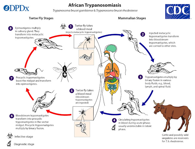
The life cycle of Trypanosoma gambiense is completed within two hosts, i.e., digenetic (Gr., di – double; genos = race), a primary vertebrate and secondary invertebrate host or vector. The vertebrate host is man and the invertebrate host is blood sucking fly, Glossina palpalis (Tsetse fly). Trypanosoma gambiense lives harmlessly in the blood of antelopes.
Part of Life Cycle in Man:
When an infected fly bites a man, it inoculates a few parasites in the blood of man. The parasites first live in the blood of the infected man, but later find their way into the cerebrospinal fluid.
While the parasites are in the blood, the infected man develops a kind of fever termed Gambia fever, but when they reach the cerebrospinal fluid, various nervous symptoms are produced in the patient leading to a lethargic condition, which has given the name sleeping sickness to the disease.
The parasites multiply by longitudinal binary fission in the blood and produce three forms of individuals, viz.,:
(i) Long and thin form’s with a free flagellum,
(ii) Short and stumpy forms with a reduced flagellum and
(iii) Intermediate forms. It has been observed that the parasites periodically increase and decrease in number in the blood of man. During the period of decrease the short and stumpy forms, which have great resisting power, survive the period of depression and the rest die. These short and stumpy forms are capable of development in the intermediate host, Glossina palpalis (Testse fly).
Part of Life Cycle in Tsetse Fly:
When a tsetse fly sucks the blood of an infected man, a number of parasites enter into the midgut of the fly along with the blood. These parasites remain in the midgut of the fly for a few days and start multiplying by longitudinal binary fission. After tenth to fifteenth day, long slender forms appear in great numbers which move forward to the proventriculus.
After several more days, the trypanosomes make their way to the fly’s salivary gland. In the salivary glands they become attached to the walls and undergo another rapid phase of multiplication by longitudinal binary fission and develop into crithidial forms.
The crithidial forms are characterised by a shorter flagellum and undulating membrane. Flagellum and undulating membrane do not extend in the hinder part of the body. Kinetoplast and basal granule are situated above the nucleus towards the anterior end.
Here the development continues for 2-5 days and the crithidial forms produce metacyclic forms (Trypanosome forms) which are now infective. These metacyclic forms pass down through the ducts and hypopharynx. When the fly bites a man, the metacyclic forms enter the blood of man along with the saliva of the fly. The whole cycle in the fly usually takes 2-30 days.
12. Transmission of Trypanosoma Gambiense:
Transmission from one vertebrate host to another is effected by an intermediate host which is a blood-sucking fly, Glossina palpaiis (Tsetse fly).
The transmission occurs in two ways:
1. Mechanical or direct transmission:
When a tsetse fly (carrier fly) bites a man infected with Trypanosome, some Trypanosomes stick to the proboscis of the fly and when the fly bites another man, the Trypanosomes are introduced into his blood, provided the time between two successive bites does not exceed 24 hours.
Such a transmission is termed mechanical or direct as the fly acts merely as a mechanical carrier and parasites do not undergo any changes in it.
2. Cyclical transmission:
When the fly sucks the blood of an infected man, the parasites along with the blood enter the mid-gut of the fly, remain there for two days and start multiplying. Parasites can be inoculated in the blood of another man only after undergoing through a set of stages. This type of transmission is known as cyclical transmission.
13. Reservoirs of Trypanosoma Gambiense:
Trypanosomes are harmless to their natural vertebrate hosts which are wild antelopes, pigs, buffaloes, etc. These wild antelopes and referred mammals are not harmed by the parasite, hence, they act as reservoir hosts from which infection is spread by the vectors or intermediate hosts.
14. Pathogenicity and Symptoms of Trypanosoma Gambiense:
The bite of an infected fly is usually followed by itching and irritation near the wound, and frequently a local dark red lesion develops. In blood, the parasite multiplies and absorbs nutrients from it. After a few days, fever and headache develop, recurring at regular intervals accompanied by increasing weakness, loss of weight and anaemia.
Usually, the parasites succeed in penetrating the lymphatic glands. Because of its infection, the lymphatic glands swell and after it the parasites enter the cerebrospinal fluid and brain causing a sleeping sickness like condition. Development of lethargic condition and recurrence of fever are the symptoms of its infection.
15. Disease Caused by Trypanosoma Gambiense:
Trypanosoma gambiense causes trypanosomiasis; most commonly referred to as sleeping sickness leading to coma stage and finally resulting into the death of the patient. In fact, two types of diseases are caused by Trypanosome which are essentially similar in symptoms. These are Gambian and Rhodesian sleeping sickness.
The Gambian sleeping sickness occurs in western part of Africa and its vector is Glossina palpalis, while Rhodesian sleeping sickness occurs in rest of Africa and its vector is Glossina morsitans. The only difference between the two is that the latter is more rapid causing the death of the patient within 3-4 months of infection.
16. Diagnosis, Treatment and Prevention of Disease Caused by Trypanosoma Gambiense:
The diagnosis is confirmed by examining fresh or stained peripheral blood or by examining the cerebrospinal fluid obtained by lumbar puncture or by examining the extract of enlarged lymphatic glands.
Treatment (Therapy):
Arsenic and antimony compounds were until recently the drugs for treatment of trypanosomiasis, but now they are rarely used except for late stages when the parasites have invaded the central nervous system.
Two drugs, Bayer 205 (also called Antrypol, Germanin or Suramin), and Pentamidine or Lomidine are now widely used for both treatment and prophylaxis of human infections. These drugs are low in toxicity, effective in treatment, and prevent reinfection for several months.
Prevention (Prophylaxis) :
The following measures are suggested for preventing the infection of this parasite:
1. By eradicating the vectors. The infection of this parasite can be checked by completely eradicating the secondary host (Tsetse fly). For this, the endemic areas should be kept clean and regular spray of insecticides like DDT is suggested which help in eradicating the fly.
2. Care should be taken to keep the reservoir hosts free from its infection.
3. Preventive medicines should be taken frequently and periodically which help to a great extent from its infection.


Comments
Post a Comment